When you wake up in the morning and look in the mirror, you probably aren’t expecting to discover something new on your face. Maybe you’re checking your skin before applying moisturizer. Maybe you’re looking for a new pimple, checking yesterday’s breakout, or simply making sure you look ready for the day. Then you notice it. A tiny white bump near your eye. A small flesh-colored lump on your cheek. A yellowish patch near your eyelid. Or perhaps a cluster of bumps that looks suspiciously like acne—but doesn’t behave like acne at all. It’s easy to assume that almost every facial bump is a pimple. After all, acne is extremely common, and clogged pores can appear seemingly overnight.
But acne is only one of many conditions that can create bumps, lumps, spots, or raised areas on the face. Your skin contains hair follicles, oil-producing glands, sweat ducts, blood vessels, connective tissue, and many other structures. Changes involving any of these structures can sometimes create something that looks like a pimple. Some bumps are tiny and white. Others are yellow, pink, brown, or the same color as the surrounding skin. Some feel completely smooth. Others feel rough or firm. Some disappear naturally, while others may remain for months or years.
Fortunately, many facial bumps are harmless.
However, appearance alone isn’t always enough to determine exactly what a bump is.
Certain benign skin growths can resemble more serious conditions, including some forms of skin cancer.
That’s why an unfamiliar bump that grows, changes, bleeds, becomes painful, repeatedly crusts over, or simply doesn’t heal deserves professional attention.
Rather than squeezing every mysterious bump like a pimple, it helps to understand some of the other possibilities.
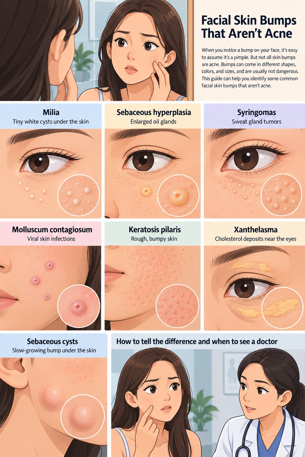
Here are several common facial skin bumps that aren’t necessarily acne—and the characteristics that can help you recognize them.
1. Milia: Tiny White Cysts Beneath the Surface
If you’ve ever noticed tiny, pearl-like white bumps around your eyes, cheeks, forehead, or nose, you may have been looking at milia.
Milia are small cysts containing keratin, a protein naturally found in the skin, hair, and nails.
They can look remarkably similar to whiteheads.
However, there’s an important difference.
A typical acne whitehead develops when a pore becomes clogged with oil, dead skin cells, and other material.
Milia are tiny keratin-filled cysts located beneath the surface of the skin.
That distinction also explains why trying to squeeze them like pimples often doesn’t work.
In fact, repeatedly picking, scratching, or squeezing the area may cause irritation, inflammation, discoloration, or even scarring.
Milia are particularly common around the eyes because the skin in that area is delicate.
They can also occur on the cheeks, nose, forehead, and elsewhere.
Sometimes they appear for no obvious reason.
In other situations, they may develop after skin injury or irritation.
Certain heavy or occlusive skin products may also contribute to milia-like bumps in some people.
The reassuring news is that milia are generally harmless.
They aren’t contagious, and they aren’t usually a sign that something is seriously wrong.
Some disappear naturally over time.
Others can remain stubbornly in place.
If you have persistent milia that bother you cosmetically, a dermatologist can evaluate them and discuss safe removal methods.
Professional extraction is generally much safer than attempting to dig them out yourself.
For prevention, gentle skin care is usually the best approach.
Avoid aggressive scrubbing around the eyes, choose products appropriate for your skin type, and be careful about using very heavy products in areas where you’re prone to developing tiny bumps.
2. Sebaceous Hyperplasia: When Oil Glands Become Enlarged
Another facial condition that can easily be mistaken for acne is sebaceous hyperplasia.
Your skin contains sebaceous glands that produce sebum, the natural oil that helps lubricate and protect your skin.
Sometimes these glands become enlarged.
When that happens, small bumps can become visible on the surface.
Sebaceous hyperplasia commonly appears on areas of the face with many oil glands, particularly the forehead, cheeks, and nose.
The bumps are often skin-colored, pale yellow, or slightly whitish.
One of their most recognizable features is a small depression or indentation in the center.
That central dip can help distinguish sebaceous hyperplasia from an ordinary pimple.
Unlike inflammatory acne, these bumps often aren’t red, swollen, or painful.
They may also persist rather than going through the familiar cycle of a pimple developing, becoming inflamed, and eventually healing.
Hormonal influences, age, genetics, and other factors may play a role in the development of sebaceous hyperplasia.
The condition is benign and generally doesn’t require treatment.
However, there’s an important reason not to diagnose it solely from photographs.
Some lesions—particularly basal cell carcinoma, a common type of skin cancer—can sometimes resemble sebaceous hyperplasia.
A dermatologist can examine a suspicious lesion more closely and determine whether additional testing is necessary.
If sebaceous hyperplasia has been professionally diagnosed and you want treatment for cosmetic reasons, a dermatologist may discuss several options.
Depending on the individual case, treatments can include procedures that destroy or remove the enlarged gland.
Some people also use ingredients such as retinoids or salicylic acid as part of their overall skin-care routine, although these shouldn’t be considered guaranteed ways of removing established lesions.
The biggest takeaway is simple:
A persistent flesh-colored bump isn’t automatically acne.
And if its appearance changes or you’re uncertain about it, getting it checked is safer than repeatedly treating it as a pimple.
3. Syringomas: Small Growths Associated With Sweat Ducts
Those tiny bumps around the eyes aren’t always milia.
Another possibility is syringomas.
Syringomas are benign growths associated with the ducts of eccrine sweat glands.
They often appear as multiple small, firm bumps.
They may be skin-colored, yellowish, tan, or slightly darker than the surrounding skin.
A classic location is around the lower eyelids and upper cheeks.
Because they often occur in groups and can be very small, people sometimes assume they’re clogged pores, milia, or acne.
However, syringomas don’t contain the same material as ordinary pimples.
They also aren’t caused by poor hygiene.
Some people develop only a few.
Others may develop numerous bumps clustered around the eyes.
Syringomas can occur in different populations and may sometimes run in families.
Certain medical or genetic conditions have also been associated with syringomas, although most people who develop ordinary syringomas don’t necessarily have an underlying disease.
The bumps themselves are benign.
They generally don’t require treatment unless their appearance bothers the person who has them.
Because the skin around the eyes is extremely delicate, trying to squeeze, cut, burn, or otherwise remove them at home can create much bigger problems than the original bumps.
Scarring and pigmentation changes can be especially noticeable in this area.
A dermatologist can first confirm whether the bumps are actually syringomas and then explain appropriate cosmetic treatment options if desired.
4. Molluscum Contagiosum: Small Bumps Caused by a Virus
Some bumps aren’t caused by clogged pores or enlarged glands at all.
Molluscum contagiosum is a viral skin infection that produces distinctive small bumps.
These bumps are usually round and firm and may appear skin-colored, pink, pearly, or occasionally slightly darker.
One of the most characteristic signs is a small indentation in the center.
At first glance, an individual molluscum lesion can resemble a pimple.
But several lesions may gradually appear in the same general area.
The surrounding skin can sometimes become red, dry, itchy, or irritated.
Molluscum contagiosum is caused by a poxvirus and can spread from person to person.
Transmission can occur through direct skin-to-skin contact.
The virus can also potentially spread through contaminated objects such as towels or clothing.
Scratching or picking the lesions can spread them to nearby areas of your own skin as well.
Molluscum is especially common among children, although adults can develop it too.
In otherwise healthy people, the infection often eventually clears as the immune system responds to the virus.
The frustrating part is that “eventually” can mean many months, and in some cases lesions can persist considerably longer.
Treatment isn’t always necessary.
However, a healthcare professional may recommend treatment in certain situations—for example, if lesions are widespread, troublesome, repeatedly irritated, or located in areas where transmission is a concern.
People with weakened immune systems should be especially cautious about persistent or extensive lesions and should discuss them with a healthcare professional.
And because molluscum is contagious, don’t assume that a cluster of unfamiliar bumps should be squeezed like ordinary pimples.
5. Keratosis Pilaris: The Rough “Chicken Skin” Texture
Some skin bumps are more noticeable by touch than by appearance.
Keratosis pilaris is one of the best examples.
People often describe the texture as “chicken skin” because affected areas feel covered in numerous tiny, rough bumps.
Keratosis pilaris most commonly appears on the upper arms and thighs.
However, it can also affect the cheeks, particularly in younger people.
The bumps develop when keratin builds up and plugs hair follicles.
Depending on skin tone and inflammation, they may look skin-colored, white, reddish, or brownish.
Keratosis pilaris often runs in families.
It’s also frequently seen in people who have dry skin or eczema.
Cold or dry weather can make the texture more noticeable because the skin becomes drier.
Fortunately, keratosis pilaris is harmless.
It’s not contagious.
And it isn’t caused by dirty skin.
Aggressive scrubbing can actually make the problem look worse by irritating the area.
Instead, gentle skin care is usually recommended.
Regular moisturization can help soften the skin.
Products containing ingredients such as lactic acid, urea, or salicylic acid may help loosen accumulated dead skin cells and improve texture for some people.
Results aren’t instant, however.
Keratosis pilaris often requires consistent skin care, and it can return when treatment is stopped.
For many people, the condition becomes less noticeable with age.
If the bumps are inflamed, severely itchy, or difficult to distinguish from another skin problem, a dermatologist can provide more specific advice.
6. Xanthelasma: Yellowish Cholesterol Deposits Around the Eyes
A yellowish bump or plaque near the eyelid deserves a different kind of attention.
Xanthelasma refers to soft, yellowish deposits that usually develop on or around the eyelids, often near the inner corners of the eyes.
They may begin as relatively small areas and become more noticeable over time.
Unlike acne, xanthelasma usually isn’t red or inflamed.
It tends to look flat or slightly raised and may have irregular borders.
These deposits contain cholesterol-rich material.
Some people who develop xanthelasma have abnormal cholesterol or other lipid levels.
Others may have normal blood lipid results.
For that reason, you shouldn’t assume that xanthelasma automatically means your cholesterol is dangerously high.
However, its appearance can be a good reason to speak with a healthcare professional.
A clinician may recommend checking cholesterol and triglyceride levels and assessing other cardiovascular risk factors depending on your health history.
The deposits themselves are usually harmless.
They don’t need to be removed unless they’re causing cosmetic concerns or other problems.
If removal is desired, several professional treatment methods may be available.
It’s important to understand that removing the visible deposit doesn’t necessarily address any underlying lipid abnormality.
Xanthelasma can also return after treatment.
That’s why the most important step isn’t simply making the bump disappear.
It’s making sure you understand why it may have developed in the first place.
7. Skin Cysts: Slow-Growing Lumps Beneath the Skin
Sometimes what appears to be an unusually large pimple is actually a cyst beneath the skin.
People frequently use the phrase “sebaceous cyst,” but many common cysts referred to by that name are actually epidermoid or pilar cysts.
These cysts can develop on areas such as the face, scalp, neck, back, and trunk.
They usually feel like a round lump underneath the skin.
Some are relatively firm.
Others feel softer.
The overlying skin may look completely normal.
Certain cysts have a small opening or dark spot in the center.
Many contain keratin rather than ordinary skin oil.
A small, uncomplicated cyst may remain almost unchanged for years.
Others slowly increase in size.
A cyst can also become inflamed.
When this happens, it may suddenly become red, swollen, tender, warm, or painful.
If it ruptures, thick material may drain from it.
This is exactly the point when many people are tempted to squeeze it.
That’s usually not a good idea.
Trying to force material out yourself can worsen inflammation, injure surrounding tissue, introduce bacteria, and potentially lead to scarring.
If a cyst is small, painless, and has been properly identified, treatment may not be necessary.
But if it becomes painful, red, rapidly enlarges, repeatedly drains, or appears infected, contact a healthcare professional.
Depending on the circumstances, a clinician may recommend monitoring it, treating inflammation or infection, or removing the cyst.
Removing only the contents doesn’t always prevent recurrence because the cyst wall may remain beneath the skin.
So, How Can You Tell Acne From Another Type of Skin Bump?
This is where things become tricky.
There isn’t one simple rule that can distinguish every pimple from every other facial bump.
Acne itself comes in several forms.
You can have blackheads.
You can have whiteheads.
You can have small inflammatory papules.
You can have pus-filled pustules.
And deeper acne can produce painful nodules or cyst-like lesions beneath the skin.
Because acne has so many different appearances, some non-acne conditions can easily be mistaken for it.
However, there are clues.
Consider How Long the Bump Has Been There
Ordinary pimples tend to change.
They may become inflamed, develop a visible head, shrink, heal, and disappear.
A tiny bump that remains almost identical for months may deserve a closer look.
That doesn’t automatically mean it’s dangerous.
Milia, syringomas, sebaceous hyperplasia, and many other benign conditions can remain stable for long periods.
The point is that persistent bumps shouldn’t automatically be treated as endless acne.
Look at the Pattern
Acne commonly affects oil-rich areas such as the forehead, nose, chin, chest, shoulders, and back.
You may see different types of acne lesions in the same area.
Some conditions have more distinctive patterns.
Milia frequently occur around the eyes and upper cheeks.
Syringomas commonly cluster around the eyelids.
Xanthelasma tends to develop around the eyelids, particularly toward the inner corners.
Keratosis pilaris creates many tiny rough bumps rather than a few isolated pimples.
Molluscum may produce multiple rounded lesions with central indentations.
Patterns can provide clues, but they still aren’t a substitute for a professional diagnosis.
Pay Attention to Texture
Run a clean finger gently across the area without squeezing.
Does the bump feel rough?
Smooth?
Firm?
Soft?
Is it beneath the skin?
Does it move slightly?
Does it feel tender?
Texture can help a dermatologist narrow down the possibilities.
For example, keratosis pilaris tends to create roughness across an area, while milia often feel like tiny hard beads beneath the skin.
Don’t Judge a Bump Only by Its Color
Red doesn’t automatically mean acne.
White doesn’t automatically mean milia.
Yellow doesn’t automatically mean xanthelasma.
Skin conditions can look different depending on a person’s natural skin tone, inflammation, lighting, and the stage of the condition.
That’s another reason online photographs should be used for general education rather than self-diagnosis.
The Biggest Mistake: Squeezing Every Bump You See
When people see something that resembles a pimple, the natural instinct is often:
“Just pop it.”
Unfortunately, that’s one of the worst approaches to an unidentified facial bump.
If the lesion isn’t acne, squeezing it may accomplish absolutely nothing.
Instead, you may create an open wound where there wasn’t one before.
Picking can cause inflammation, bleeding, infection, scarring, and post-inflammatory pigmentation.
This is particularly important around the eyes, where the skin is thin and delicate.
If something has been sitting on your face for weeks or months and refuses to behave like a normal pimple, stop trying to force it to behave like one.
Get it identified.
When Should You See a Doctor or Dermatologist?
Most facial bumps turn out to be benign.
Still, there are situations when waiting and watching isn’t the best approach.
Make an appointment with a healthcare professional if you notice a bump that:
- Continues growing.
- Changes shape or color.
- Becomes persistently painful or tender.
- Bleeds without an obvious injury.
- Repeatedly crusts, heals, and returns.
- Develops an open sore that doesn’t heal.
- Becomes very red, hot, swollen, or produces drainage.
- Appears suddenly in large numbers.
- Spreads rapidly.
- Looks noticeably different from your other spots or moles.
- Persists and you simply can’t identify what it is.
You also don’t have to wait for a lesion to become frightening before seeing a dermatologist.
If something is bothering you, changing your appearance, or making you anxious because you don’t know what it is, that’s a reasonable reason to ask a professional.
Why Self-Diagnosing From Photos Can Be Difficult
Social media has made it easier than ever to search for pictures of skin conditions.
You notice a bump.
You search for “tiny white bump under eye.”
Five minutes later, you’ve looked at photographs of milia, syringomas, clogged pores, skin cancer, warts, cysts, and several conditions you’ve never heard of before.
Instead of feeling reassured, you’re more confused.
There’s a reason for that.
Many skin conditions overlap in appearance.
Lighting can change how a lesion looks in a photograph.
Different skin tones can also change the visible signs of inflammation.
And a two-dimensional photograph can’t reveal important information about texture, firmness, depth, or how a lesion behaves when examined closely.
Dermatologists may use magnification and specialized examination techniques.
If the diagnosis remains uncertain, they may recommend a biopsy or another test.
That’s why an online article can help you understand possibilities—but it can’t tell you with certainty what a specific bump on your face is.
Your Skin Is Constantly Changing
Our skin isn’t static.
It responds to age, hormones, weather, sunlight, medications, cosmetics, genetics, immune activity, and many other influences.
Something you never experienced in your twenties may suddenly appear later in life.
A product you’ve used for years may no longer suit your skin.
A harmless bump may appear without any obvious explanation.
This doesn’t mean you need to panic every time your face changes.
It means paying attention is useful.
You see your own face more often than almost anyone else does.
That gives you an advantage.
You may be the first person to notice when something appears, grows, changes, or refuses to heal.
A Simple Approach When You Discover an Unfamiliar Bump
First, resist the temptation to squeeze it.
Take note of where it is and what it looks like.
If necessary, take a clear photograph for your own records so you can compare it later.
Continue gentle skin care.
Avoid aggressively scrubbing or applying multiple harsh treatments at once.
Watch for changes.
If the bump disappears, it may have been a temporary irritation or acne lesion.
If it stays exactly the same, that information is useful too.
And if it grows, changes, bleeds, becomes painful, or worries you, have it professionally evaluated.
Final Thoughts
Finding a strange bump on your face can be unsettling, especially when it appears somewhere highly visible.
But not every bump is acne.
And not every non-acne bump is something dangerous.
Milia can create tiny white cysts.
Sebaceous hyperplasia can cause enlarged oil glands to become visible.
Syringomas can produce clusters of small bumps around the eyes.
Molluscum contagiosum can create viral, dimpled lesions.
Keratosis pilaris can make the skin feel rough and bumpy.
Xanthelasma can cause yellowish deposits around the eyelids.
And common skin cysts can create slow-growing lumps beneath the surface.
Many of these conditions are harmless.
The challenge is that different skin conditions can sometimes look remarkably similar.
That’s why the goal shouldn’t be to diagnose yourself perfectly after looking at a few photographs.
The goal is to recognize when something doesn’t behave like ordinary acne and know when it’s time to get professional advice.
Pay attention to your skin.
Treat it gently.
Don’t squeeze unfamiliar growths.
And remember that a persistent or changing bump deserves more attention than another layer of acne cream.
Your mirror can help you notice a change.
But when you’re uncertain about what that change means, a qualified healthcare professional or dermatologist is the person who can give you the diagnosis you actually need.
This article is for general educational purposes and isn’t a substitute for diagnosis or treatment from a qualified healthcare professional.